HLA-G
| HLA-G | |||||||||||||||||||||||||||||||||||||||||||||||||||
|---|---|---|---|---|---|---|---|---|---|---|---|---|---|---|---|---|---|---|---|---|---|---|---|---|---|---|---|---|---|---|---|---|---|---|---|---|---|---|---|---|---|---|---|---|---|---|---|---|---|---|---|
 | |||||||||||||||||||||||||||||||||||||||||||||||||||
| Identifiers | |||||||||||||||||||||||||||||||||||||||||||||||||||
| Aliases | HLA-G, MHC-G, major histocompatibility complex, class I, G | ||||||||||||||||||||||||||||||||||||||||||||||||||
| External IDs | OMIM: 142871; MGI: 95915; HomoloGene: 133255; GeneCards: HLA-G; OMA:HLA-G - orthologs | ||||||||||||||||||||||||||||||||||||||||||||||||||
| |||||||||||||||||||||||||||||||||||||||||||||||||||
| |||||||||||||||||||||||||||||||||||||||||||||||||||
| |||||||||||||||||||||||||||||||||||||||||||||||||||
| |||||||||||||||||||||||||||||||||||||||||||||||||||
| |||||||||||||||||||||||||||||||||||||||||||||||||||
| Wikidata | |||||||||||||||||||||||||||||||||||||||||||||||||||
| |||||||||||||||||||||||||||||||||||||||||||||||||||


HLA-G histocompatibility antigen, class I, G, also known as human leukocyte antigen G (HLA-G), is a protein that in humans is encoded by the HLA-G gene.[5]
HLA-G belongs to the HLA nonclassical class I heavy chain paralogues. Classical HLA I proteins are found on all nucleated cells and express peptides in their peptide binding groove. They can express "self" peptides when the cell is healthy as well as foreign peptides when the cell is infected by a parasite or cancer. HLA-G is a nonclassical protein and serves a different function from classical HLA class I molecules, but it still expresses a nine amino acid peptide in its peptide binding groove.[6] The third and ninth amino acid in the peptide sequence serve as anchor residues, and are thus conserved in all the peptides HLA-G bind to.
Structure
This class I molecule is a heterodimer consisting of a heavy chain and a light chain (beta-2 microglobulin). The heavy chain is anchored in the membrane. HLA-G is coded for by 88 alleles.[7] The heavy chain is approximately 45 kDa and its gene contains 8 exons. Exon one encodes the leader peptide, exons 2 and 3 encode the alpha1 and alpha2 domain, which both bind the peptide, exon 4 encodes the alpha3 domain, exon 5 encodes the transmembrane region, and exon 6 encodes the cytoplasmic tail.[5] Exon 7 and 8 are not translated due to a stop codon present in exon 6.[8]
HLA-G can be expressed under at least seven isoforms through alternative splicing, called HLA-G1, HLA-G2,..., HLA-G7.[6][9] The protein can be both membrane-bound and soluble. HLA-G1 through G4 are membrane bound and HLA-G5 through G7 are soluble.[6] HLA-G1 and HLA-G5 are the most studied isoforms due to the wider availability of antibodies targeting them. HLA-G can present a more narrow variety of peptides than its classical HLA class I counterparts due to it having a more limited polymorphism.
Function
In the Human Body
HLA-G is a major immune checkpoint, meaning it downregulates the immune system's response.[9] Soluble HLA-G can be found in the saliva, ascitic fluid, plasma, thymus, seminal plasma, cerebrospinal fluid, and in first and second term placentas.[10] Membrane-bound HLA-G is predominantly found on trophoblast cells in the placenta, but it is also found in the thymus, cornea, erythroblasts, and mesenchymal stem cells.[7] It can be upregulated in cancers.[9] Peptides are connected to HLA-G by the peptide loading complex in the endoplasmic reticulum.[6]
Pregnancy
HLA-G plays a role in immune tolerance in pregnancy, being expressed in the placenta by extravillous trophoblast cells (EVT), while the classical MHC class I genes (HLA-A and HLA-B) are not.[6][11] As HLA-G was first identified in placenta samples, many studies have evaluated its role in pregnancy disorders, such as preeclampsia and recurrent pregnancy loss.[12] Its downregulation is related to HLA-A and -B downregulation results in protection from cytotoxic T cell responses, but would in theory result in a missing self response by natural killer cells. HLA-G is a ligand for natural killer (NK) cell inhibitory receptor KIR2DL4, and therefore expression of this HLA by the trophoblast defends it against NK cell-mediated death.[13]
The presence of soluble HLA-G (sHLA-G) in embryos is associated with better pregnancy rates. In order to optimize pregnancy rates, there is significant evidence that a morphological scoring system is the best strategy for the selection of embryos.[14] However, presence of soluble HLA-G might be considered as a second parameter if a choice has to be made between embryos of morphologically equal quality.[14]
Parasitic Infections
HLA-G has been shown to modulate the body's response to parasitic diseases. Recent studies have emerged suggesting a link between HLA-G and P. falciparum, which is one of the most dangerous malaria strains.[7] In pregnant women, P. falciparum can infect the placenta, causing low birth weights and other complications. High levels of soluble HLA-G have been linked to higher instances of low birth weights. There is also a link between HLA-G expression and Human African trypanosomiasis (HAT).[7] People with higher levels of soluble HLA-G are more likely to be diagnosed with the disease. There may also be genetic differences driving the instance and severity of HAT, as a few single nucleotide polymorphisms have been associated with higher levels of HAT. There is also an effect in Toxoplasmosis infections in pregnant women, where HLA-G is upregulated to protect the fetus from inflammation.[7] Treatment of cells with IL-10 leads to a downregulation of HLA-G, which could be an avenue for therapy in instances where too much HLA-G is produced. Individuals with Visceral leishmaniasis infections also have higher levels of soluble HLA-G, which may be due to a strategy by Leishmania to evade the immune system.[7]
Cancer
HLA-G has been shown to be associated with tumor escape in cancers, because it causes the immune system to not pay attention to cancer cells. Because it is upregulated in cancer cells, it could serve as a potential target for immunotherapy.[9] Monoclonal antibodies that bind to HLA-G have been used successfully against cancers as part of a strategy to inhibit immune checkpoints.[6] HLA-G has potential utility as a tumor marker due to the large increase in HLA-G in many cancers, including breast cancer, ovarian cancer, and lung cancer.[10] Increased expression of HLA-G has been associated with the metastatic potential of tumor cells.[15]
Allergy
HLA-G has links to allergenic responses in the body. Soluble HLA-G levels are higher in the serum of people with allergic rhinitis, or hay fever.[16] Additionally, single nucleotide polymorphisms in HLA-G have been connected to an increased likelihood of having asthma. Papillary cells expressing HLA-G were found in patients with atopic dermatitis.[16]
Interactions
HLA-G has been shown to interact with CD8A.[17][18] When in its soluble form, HLA-G interacts with Ig-like transcript 2 (ILT2), a leukocyte receptor. When it’s membrane bound, it interacts with Ig-like transcript 4 (ILT4).[6][7] Soluble HLA-G can bind to KIR2DL4, which is often found on the surface of natural killer cells. The identity of the peptide presented by HLA-G is unrelated to the binding of HLA with KIR2DL4, ILT2, or ILT4.[6] Because HLA-G interacts with receptors using a variety of its domains, multiple antibodies are necessary to inhibit all of its functions.
Both ILT2 and ILT4 cause negative intracellular signaling.[7] In monocytes, binding to either ILT2 or ILT4 receptors cause the inhibition of monocyte/macrophage mediated toxicity. In dendritic cells, binding to both receptors can prevent dendritic cells from maturing and prevent the activation of T cells.[7] Additionally, HLA-G may interact with ILT4 receptors on the surface of neutrophils to inhibit phagocytosis. In natural killer cells, HLA-G binds with the ILT2 receptor to inhibit the secretion of IFN-γ, a cytokine that can activate macrophages and stimulate natural killer cells and neutrophils.[7] HLA-G binds to ILT2 on B cells to cause the inhibition of B cell proliferation, differentiation, and the secretion of antibodies. It binds to ILT2 on T cells to downregulate T cell chemokine expression. The cytokine expression of T cells mimics that of TH2 cells. HLA-G causes apoptosis in CD8+ T cells.[6] All together these effects serve to decrease the inflammatory response of the immune system.
References
- ^ a b c ENSG00000233095, ENSG00000237216, ENSG00000276051, ENSG00000204632, ENSG00000235346, ENSG00000235680, ENSG00000206506 GRCh38: Ensembl release 89: ENSG00000230413, ENSG00000233095, ENSG00000237216, ENSG00000276051, ENSG00000204632, ENSG00000235346, ENSG00000235680, ENSG00000206506 – Ensembl, May 2017
- ^ a b c GRCm38: Ensembl release 89: ENSMUSG00000016206 – Ensembl, May 2017
- ^ "Human PubMed Reference:". National Center for Biotechnology Information, U.S. National Library of Medicine.
- ^ "Mouse PubMed Reference:". National Center for Biotechnology Information, U.S. National Library of Medicine.
- ^ a b "Entrez Gene: HLA-G HLA-G histocompatibility antigen, class I, G".
- ^ a b c d e f g h i Attia JV, Dessens CE, van de Water R, Houvast RD, Kuppen PJ, Krijgsman D (November 2020). "The Molecular and Functional Characteristics of HLA-G and the Interaction with Its Receptors: Where to Intervene for Cancer Immunotherapy?". International Journal of Molecular Sciences. 21 (22): 8678. doi:10.3390/ijms21228678. PMC 7698525. PMID 33213057.
- ^ a b c d e f g h i j Zhuang B, Shang J, Yao Y (2021). "HLA-G: An Important Mediator of Maternal-Fetal Immune-Tolerance". Frontiers in Immunology. 12: 744324. doi:10.3389/fimmu.2021.744324. PMC 8586502. PMID 34777357.
- ^ Castelli EC, Mendes-Junior CT, Veiga-Castelli LC, Roger M, Moreau P, Donadi EA (November 2011). "A comprehensive study of polymorphic sites along the HLA-G gene: implication for gene regulation and evolution". Molecular Biology and Evolution. 28 (11): 3069–3086. doi:10.1093/molbev/msr138. PMID 21622995.
- ^ a b c d Loustau M, Anna F, Dréan R, Lecomte M, Langlade-Demoyen P, Caumartin J (2020). "HLA-G Neo-Expression on Tumors". Frontiers in Immunology. 11: 1685. doi:10.3389/fimmu.2020.01685. PMC 7456902. PMID 32922387.
- ^ a b Li P, Wang N, Zhang Y, Wang C, Du L (2021). "HLA-G/sHLA-G and HLA-G-Bearing Extracellular Vesicles in Cancers: Potential Role as Biomarkers". Frontiers in Immunology. 12: 791535. doi:10.3389/fimmu.2021.791535. PMC 8636042. PMID 34868081.
- ^ Jay Iams; Creasy, Robert K.; Resnik, Robert; Robert Reznik (2004). Maternal-fetal medicine. Philadelphia: W.B. Saunders Co. pp. 31–32. ISBN 978-0-7216-0004-8.
- ^ Michita RT, Zambra FM, Fraga LR, Sanseverino MT, Callegari-Jacques SM, Vianna P, Chies JA (October 2016). "A tug-of-war between tolerance and rejection - New evidence for 3'UTR HLA-G haplotypes influence in recurrent pregnancy loss". Human Immunology. 77 (10): 892–897. doi:10.1016/j.humimm.2016.07.004. PMID 27397898.
- ^ Lash GE, Robson SC, Bulmer JN (March 2010). "Review: Functional role of uterine natural killer (uNK) cells in human early pregnancy decidua". Placenta. 31 (Suppl): S87–S92. doi:10.1016/j.placenta.2009.12.022. PMID 20061017.
- ^ a b Rebmann V, Switala M, Eue I, Grosse-Wilde H (July 2010). "Soluble HLA-G is an independent factor for the prediction of pregnancy outcome after ART: a German multi-centre study". Human Reproduction. 25 (7): 1691–1698. doi:10.1093/humrep/deq120. PMID 20488801.
- ^ Bassey-Archibong BI, Rajendra Chokshi C, Aghaei N, Kieliszek AM, Tatari N, McKenna D, et al. (February 2023). "An HLA-G/SPAG9/STAT3 axis promotes brain metastases". Proceedings of the National Academy of Sciences of the United States of America. 120 (8): e2205247120. Bibcode:2023PNAS..12005247B. doi:10.1073/pnas.2205247120. PMC 9974476. PMID 36780531.
- ^ a b Negrini S, Contini P, Murdaca G, Puppo F (2022). "HLA-G in Allergy: Does It Play an Immunoregulatory Role?". Frontiers in Immunology. 12: 789684. doi:10.3389/fimmu.2021.789684. PMC 8784385. PMID 35082780.
- ^ Gao GF, Willcox BE, Wyer JR, Boulter JM, O'Callaghan CA, Maenaka K, et al. (May 2000). "Classical and nonclassical class I major histocompatibility complex molecules exhibit subtle conformational differences that affect binding to CD8alphaalpha". The Journal of Biological Chemistry. 275 (20): 15232–15238. doi:10.1074/jbc.275.20.15232. PMID 10809759.
- ^ Sanders SK, Giblin PA, Kavathas P (September 1991). "Cell-cell adhesion mediated by CD8 and human histocompatibility leukocyte antigen G, a nonclassical major histocompatibility complex class 1 molecule on cytotrophoblasts". The Journal of Experimental Medicine. 174 (3): 737–740. doi:10.1084/jem.174.3.737. PMC 2118947. PMID 1908512.
Further reading
- Carosella ED, Favier B, Rouas-Freiss N, Moreau P, Lemaoult J (May 2008). "Beyond the increasing complexity of the immunomodulatory HLA-G molecule". Blood. 111 (10): 4862–4870. doi:10.1182/blood-2007-12-127662. PMID 18334671. S2CID 19170578.
- Carosella ED, Moreau P, Lemaoult J, Rouas-Freiss N (March 2008). "HLA-G: from biology to clinical benefits". Trends in Immunology. 29 (3): 125–132. doi:10.1016/j.it.2007.11.005. PMID 18249584.
- Arnaiz-Villena A, Martinez-Laso J, Alvarez M, Castro MJ, Varela P, Gomez-Casado E, et al. (1997). "Primate Mhc-E and -G alleles". Immunogenetics. 46 (4): 251–266. doi:10.1007/s002510050271. PMID 9218527. S2CID 2918451.
- Le Bouteiller P (February 2000). "HLA-G in the human placenta: expression and potential functions". Biochemical Society Transactions. 28 (2): 208–212. doi:10.1042/bst0280208. PMID 10816129.
- Geyer M, Fackler OT, Peterlin BM (July 2001). "Structure--function relationships in HIV-1 Nef". EMBO Reports. 2 (7): 580–585. doi:10.1093/embo-reports/kve141. PMC 1083955. PMID 11463741.
- Langat DK, Hunt JS (November 2002). "Do nonhuman primates comprise appropriate experimental models for studying the function of human leukocyte antigen-G?". Biology of Reproduction. 67 (5): 1367–1374. doi:10.1095/biolreprod.102.005587. PMID 12390864.
- Moreau P, Dausset J, Carosella ED, Rouas-Freiss N (November 2002). "Viewpoint on the functionality of the human leukocyte antigen-G null allele at the fetal-maternal interface". Biology of Reproduction. 67 (5): 1375–1378. doi:10.1095/biolreprod.102.005439. PMID 12390865.
- Moreau P, Rousseau P, Rouas-Freiss N, Le Discorde M, Dausset J, Carosella ED (September 2002). "HLA-G protein processing and transport to the cell surface". Cellular and Molecular Life Sciences. 59 (9): 1460–1466. doi:10.1007/s00018-002-8521-8. PMID 12440768. S2CID 9352496.
- Greenway AL, Holloway G, McPhee DA, Ellis P, Cornall A, Lidman M (April 2003). "HIV-1 Nef control of cell signalling molecules: multiple strategies to promote virus replication". Journal of Biosciences. 28 (3): 323–335. doi:10.1007/BF02970151. PMID 12734410. S2CID 33749514.
- Bénichou S, Benmerah A (January 2003). "[The HIV nef and the Kaposi-sarcoma-associated virus K3/K5 proteins: "parasites"of the endocytosis pathway]". Médecine/Sciences. 19 (1): 100–106. doi:10.1051/medsci/2003191100. PMID 12836198.
- Le Bouteiller P, Legrand-Abravanel F, Solier C (April 2003). "Soluble HLA-G1 at the materno-foetal interface--a review". Placenta. 24 (Suppl A): S10–S15. doi:10.1053/plac.2002.0931. PMID 12842408.
- Sköld M, Behar SM (October 2003). "Role of CD1d-restricted NKT cells in microbial immunity". Infection and Immunity. 71 (10): 5447–5455. doi:10.1128/IAI.71.10.5447-5455.2003. PMC 201095. PMID 14500461.
- Wiendl H, Mitsdoerffer M, Weller M (November 2003). "Express and protect yourself: the potential role of HLA-G on muscle cells and in inflammatory myopathies". Human Immunology. 64 (11): 1050–1056. doi:10.1016/j.humimm.2003.07.001. PMID 14602235.
- Urosevic M, Dummer R (November 2003). "HLA-G in skin cancer: a wolf in sheep's clothing?". Human Immunology. 64 (11): 1073–1080. doi:10.1016/j.humimm.2003.08.351. PMID 14602238.
- Carosella ED, Moreau P, Le Maoult J, et al. (2003). HLA-G Molecules: From Maternal–Fetal Tolerance to Tissue Acceptance. Advances in Immunology. Vol. 81. pp. 199–252. doi:10.1016/S0065-2776(03)81006-4. ISBN 978-0-12-022481-4. PMID 14711057.
- Leavitt SA, SchOn A, Klein JC, Manjappara U, Chaiken IM, Freire E (February 2004). "Interactions of HIV-1 proteins gp120 and Nef with cellular partners define a novel allosteric paradigm". Current Protein & Peptide Science. 5 (1): 1–8. doi:10.2174/1389203043486955. PMID 14965316.
- Le Maoult J, Rouas-Freiss N, Le Discorde M, Moreau P, Carosella ED (March 2004). "[HLA-G in organ transplantation]". Pathologie-Biologie. 52 (2): 97–103. doi:10.1016/j.patbio.2003.04.006. PMID 15001239.
- Tolstrup M, Ostergaard L, Laursen AL, Pedersen SF, Duch M (April 2004). "HIV/SIV escape from immune surveillance: focus on Nef". Current HIV Research. 2 (2): 141–151. doi:10.2174/1570162043484924. PMID 15078178.
- Joseph AM, Kumar M, Mitra D (January 2005). "Nef: "necessary and enforcing factor" in HIV infection". Current HIV Research. 3 (1): 87–94. doi:10.2174/1570162052773013. PMID 15638726.
- McIntire RH, Hunt JS (April 2005). "Antigen presenting cells and HLA-G--a review". Placenta. 26 (Suppl A): S104–S109. doi:10.1016/j.placenta.2005.01.006. PMID 15837058.
- Anderson JL, Hope TJ (April 2004). "HIV accessory proteins and surviving the host cell". Current HIV/AIDS Reports. 1 (1): 47–53. doi:10.1007/s11904-004-0007-x. PMID 16091223. S2CID 34731265.
- v
- t
- e
PDB gallery
-
 1ydp: 1.9A crystal structure of HLA-G
 -
 2d31: Crystal structure of disulfide-linked HLA-G dimer
 -
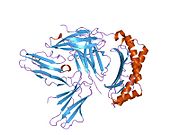 2dyp: Crystal Structure of LILRB2(LIR2/ILT4/CD85d) complexed with HLA-G


