Vascular endothelial growth factor A
Protein involved in blood vessel growth
| VEGFA | |||||||||||||||||||||||||||||||||||||||||||||||||||
|---|---|---|---|---|---|---|---|---|---|---|---|---|---|---|---|---|---|---|---|---|---|---|---|---|---|---|---|---|---|---|---|---|---|---|---|---|---|---|---|---|---|---|---|---|---|---|---|---|---|---|---|
 | |||||||||||||||||||||||||||||||||||||||||||||||||||
| Identifiers | |||||||||||||||||||||||||||||||||||||||||||||||||||
| Aliases | VEGFA, MVCD1, VEGF, VPF, vascular endothelial growth factor A | ||||||||||||||||||||||||||||||||||||||||||||||||||
| External IDs | OMIM: 192240; MGI: 103178; HomoloGene: 2534; GeneCards: VEGFA; OMA:VEGFA - orthologs | ||||||||||||||||||||||||||||||||||||||||||||||||||
| |||||||||||||||||||||||||||||||||||||||||||||||||||
| |||||||||||||||||||||||||||||||||||||||||||||||||||
| |||||||||||||||||||||||||||||||||||||||||||||||||||
| |||||||||||||||||||||||||||||||||||||||||||||||||||
| |||||||||||||||||||||||||||||||||||||||||||||||||||
| Wikidata | |||||||||||||||||||||||||||||||||||||||||||||||||||
| |||||||||||||||||||||||||||||||||||||||||||||||||||


Vascular endothelial growth factor A (VEGF-A) is a protein that in humans is encoded by the VEGFA gene.[5]
Function
This gene is a member of the platelet-derived growth factor (PDGF)/vascular endothelial growth factor (VEGF) family and encodes a protein that is often found as a disulfide linked homodimer. This protein is a glycosylated mitogen that specifically acts on endothelial cells and has various effects, including mediating increased vascular permeability, inducing angiogenesis, vasculogenesis, and endothelial cell growth, promoting cell migration, and inhibiting apoptosis. Alternatively, spliced transcript variants, encoding either freely secreted or cell-associated isoforms, have been characterized.[6]
VEGF-A shows prominent activity with vascular endothelial cells, primarily through its interactions with the VEGFR1 and -R2 receptors found prominently on the endothelial cell membrane. However, it does have effects on a number of other cell types (e.g., stimulation monocyte/macrophage migration, neurons, cancer cells, kidney and epithelial cells ). In vitro, VEGF-A has been shown to stimulate endothelial cell mitogenesis and cell migration. VEGF-A is also a vasodilator and increases microvascular permeability and was originally referred to as vascular permeability factor.
During embryonic development angiogenesis is initiated as mesoderm mesenchyme cells are specified to differentiate into angioblasts, expressing the Vascular Endothelial Growth Factor Receptor (VEGFR-2). As embryonic tissue utilizes more oxygen than it receives from diffusion, it becomes hypoxic. These cells will secrete the signaling molecule vascular endothelial factor A (VEGFA) which will recruit the angioblasts expressing its partnering receptor to the site of future angiogenesis. The angioblasts will create scaffolding structures that form the primary capillary plexus from where the local vasculature system will develop. Disruption of this gene in mice resulted in abnormal embryonic blood vessel formation, resulting in underdeveloped vascular structures. This gene is also upregulated in many tumors and its expression is correlated with tumor development and is a target in many developing cancer therapeutics. Elevated levels of this protein are found in patients with POEMS syndrome, also known as Crow-Fukase syndrome which is a hemangioblastic proliferative disorder. Allelic variants of this gene have been associated with microvascular complications of diabetes 1 and atherosclerosis.
VEGF-A Overview
Vascular endothelial growth factor A (VEGF-A) is a dimeric glycoprotein that plays a significant role in neurons and is considered to be the main, dominant inducer of the growth of blood vessels. VEGFA is essential for adults during organ remodeling and diseases that involve blood vessels, for example, in wound healing, tumor angiogenesis, diabetic retinopathy, and age-related macular degeneration. During early vertebrate development, vasculogenesis occurs which means that the endothelial condense into the blood vessels. The differentiation of endothelial cells is dependent upon the expression of VEGFA and if the expression is abolished then it can result in the death of the embryo. VEGFA is produced by a group of three major isoforms as a result of alternative splicing and if any three isoforms are produced (VEGFA120, VEGFA164, and VEGFA188) then this will not result in vessel defects and death of the full VEGFA knockout in mice. VEGFA is essential in the role of neurons because they too need vascular supply and abolishing the expression of VEGFA from neural progenitors will result in defects of the brain vascularization and neuronal apoptosis. Anti-VEGFA therapy can be used to treat patients with undesirable angiogenesis and vascular leakage in cancer and eye diseases but also could result in the inhibition of neurogenesis and neuroprotection. VEGFA could be used to treat patients with neurodegenerative and neuropathic conditions and also increase vascular permeability which will stop the blood-brain barrier and increase inflammatory cell infiltration.[7][8][9]
Usage
- Angiogenesis
- ↑ Migration of endothelial cells
- ↑ mitosis of endothelial cells
- ↑ Matrix metalloproteinase activity
- ↑ αvβ3 activity
- creation of blood vessel lumen
- creates lumen
- creates fenestrations
- Chemotactic for macrophages and granulocytes
- Vasodilation (indirectly by NO release)
Also tumour suppression.[10]
Clinical significance
Elevated levels of this protein is linked to POEMS syndrome, also known as Crow-Fukase syndrome.[11] Mutations in this gene have been associated with proliferative and nonproliferative diabetic retinopathy.[12]
Treatment of ischemic heart disease
In ischemic cardiomyopathy, blood flow to the muscle cells of the heart is either partially or completely reduced, leading to cell death and scar tissue formation. Because the muscle cells are replaced with fibrous tissue, the heart loses its ability to contract, compromising heart function.[13] Normally, if blood flow to the heart is compromised, over time, new blood vessels will develop, providing alternative circulation to the affected cells. The viability of the heart following severely restricted blood flow is dependent on the ability of the heart to provide this collateral circulation.[14] Expression of VEGF-A has been found to be induced by myocardial ischemia and a higher level of expression of VEGF-A has been associated with better collateral circulation development during ischemia.[15][16]
VEGF-A activation
When cells are deprived of oxygen, they increase their production of VEGF-A. VEGF-A mediates the growth of new blood vessels from pre-existing vessels (angiogenesis) by binding to the cell surface receptors VEGFR1 and VEGFR2, two tyrosine kinases located in endothelial cells of the cardiovascular system. These two receptors act through different pathways to contribute to endothelial cell proliferation and migration, and formation of tubular structures.[17]
VEGFR2
The binding of VEGF-A to VEGFR2 causes two VEGFR2 molecules to combine to form a dimer. Following this dimerization, through the action of the receptor itself, a phosphate group is added to certain tyrosines within the molecule in a process called auto-phosphorylation.[18] The autophosphorylation of these amino acids allows for signalling molecules within to the cell to bind to the receptor and become activated. These signalling molecules include VEGF-receptor activated protein (VRAP), PLC- γ and Nck.[19][20][21]
Each of these is important in the signalling required for angiogenesis. VRAP (also known as T-cell specific adaptor) and Nck signalling are important in reorganization of the structural components of the cell, allowing for cells to move around to areas where they are needed.[21][22] PLC- γ is vital to the proliferative effects of VEGF-A signalling. Activation of the phospholipase PLC- γ results in an increase in calcium levels in the cell, leading to the activation of protein kinase C (PKC).[23] PKC phosphorylates the mitogen-activated protein kinase (MAPK) ERK which then moves to the nucleus of the cell and takes part in nuclear signalling.[24] Once in the nucleus, ERK activates various transcription factors which initiate expression of genes involved in cellular proliferation.[25] Activation of a different MAPK (p38 MAPK) by VEGFR2 is important in the transcription of genes associated with cellular migration.[26]
VEGFR1
The tyrosine kinase activity of VEGFR1 is less efficient than that of VEGFR2 and its activation alone is insufficient to bring about the proliferative effects of VEGF-A.[27] The major role of VEGFR1 is to recruit the cells responsible in blood cell development.[28]
Current research
It has been shown that injection of VEGF-A in dogs following severely restricted blood flow to the heart caused an increase in collateral blood vessel formation compared to the dogs who did not receive the VEGF-A treatment.[16] It was also shown in dogs that delivery of VEGF-A to areas of the heart with little or no blood flow enhanced collateral blood vessel formation and increased the viability of the cells in that area.[29] In gene therapy, DNA which encodes the gene of interest is integrated into a vector along with elements that are able to promote the gene's expression. The vector is then injected either into muscle cells of the heart or the blood vessels supplying the heart. The natural machinery of the cell is then used to express these genes.[30] Currently, human clinical trials are being conducted to study the effectiveness of gene therapy with VEGF-A in restoring blood flow and function to areas of the heart that have severely restricted blood flow.[31][32][33] So far, this type of therapy has proven both safe and beneficial.[33][34]
Interactions
Vascular endothelial growth factor A has been shown to interact with:
See also
- Vascular endothelial growth factor
- Bevacizumab (or Avastin) anti VEGF-A human antibody drug.
References
- ^ a b c GRCh38: Ensembl release 89: ENSG00000112715 – Ensembl, May 2017
- ^ a b c GRCm38: Ensembl release 89: ENSMUSG00000023951 – Ensembl, May 2017
- ^ "Human PubMed Reference:". National Center for Biotechnology Information, U.S. National Library of Medicine.
- ^ "Mouse PubMed Reference:". National Center for Biotechnology Information, U.S. National Library of Medicine.
- ^ Mattei MG, Borg JP, Rosnet O, Marmé D, Birnbaum D (February 1996). "Assignment of vascular endothelial growth factor (VEGF) and placenta growth factor (PLGF) genes to human chromosome 6p12-p21 and 14q24-q31 regions, respectively". Genomics. 32 (1): 168–169. doi:10.1006/geno.1996.0098. PMID 8786112.
- ^ "Entrez Gene: Vascular endothelial growth factor A".
- ^ Mackenzie, Francesca, and Ruhrberg, Christiana. "Diverse Roles for VEGF-A in the Nervous System." Development (n.d.): 1371-380. http://dev.biologist.org/. 15 Apr. 2012. Web. 19 Mar. 2013.
- ^ Creuzet, Sophie, Gérard Couly, Christine Vincent, and Nicole M. Douarin. "Negative Effect of Hox Gene Expression on the Development of the Neural." Development (n.d.): 4301-313. http://dev.biologists.org/. 15 Sept. 2002. Web. 19 Mar. 2013.
- ^ United States of America. Johns Hopkins University. http://omim.org. By Victor A. McKusick. Johns Hopkins University, 26 Feb. 2013. Web. 19 Mar. 2013.
- ^ Stockmann C, Doedens A, Weidemann A, Zhang N, Takeda N, Greenberg JI, Cheresh DA, Johnson RS (November 2008). "Deletion of vascular endothelial growth factor in myeloid cells accelerates tumorigenesis". Nature. 456 (7223): 814–818. Bibcode:2008Natur.456..814S. doi:10.1038/nature07445. PMC 3103772. PMID 18997773.
- ^ Dispenzieri A (November 2007). "POEMS syndrome". Blood Rev. 21 (6): 285–299. doi:10.1016/j.blre.2007.07.004. PMID 17850941.
- ^ Watanabe D, Suzuma K, Suzuma I, Ohashi H, Ojima T, Kurimoto M, Murakami T, Kimura T, Takagi H (March 2005). "Vitreous levels of angiopoietin 2 and vascular endothelial growth factor in patients with proliferative diabetic retinopathy". Am. J. Ophthalmol. 139 (3): 476–481. doi:10.1016/j.ajo.2004.10.004. PMID 15767056.
- ^ Beltrami CA, Finato N, Rocco M, Feruglio GA, Puricelli C, Cigola E, Quaini F, Sonnenblick EH, Olivetti G, Anversa P (January 1994). "Structural basis of end-stage failure in ischemic cardiomyopathy in humans". Circulation. 89 (1): 151–63. doi:10.1161/01.cir.89.1.151. PMID 8281642.
- ^ Sabia PJ, Powers ER, Ragosta M, Sarembock IJ, Burwell LR, Kaul S (December 1992). "An association between collateral blood flow and myocardial viability in patients with recent myocardial infarction". N. Engl. J. Med. 327 (26): 1825–1831. doi:10.1056/NEJM199212243272601. PMID 1448120.
- ^ Banai S, Shweiki D, Pinson A, Chandra M, Lazarovici G, Keshet E (August 1994). "Upregulation of vascular endothelial growth factor expression induced by myocardial ischaemia: implications for coronary angiogenesis". Cardiovasc. Res. 28 (8): 1176–9. doi:10.1093/cvr/28.8.1176. PMID 7525061.
- ^ a b Chalothorn D, Clayton JA, Zhang H, Pomp D, Faber JE (July 2007). "Collateral density, remodeling, and VEGF-A expression differ widely between mouse strains". Physiol. Genomics. 30 (2): 179–191. CiteSeerX 10.1.1.325.6534. doi:10.1152/physiolgenomics.00047.2007. PMID 17426116.
- ^ Huusko J, Merentie M, Dijkstra MH, Ryhänen MM, Karvinen H, Rissanen TT, Vanwildemeersch M, Hedman M, Lipponen J, Heinonen SE, Eriksson U, Shibuya M, Ylä-Herttuala S (April 2010). "The effects of VEGF-R1 and VEGF-R2 ligands on angiogenic responses and left ventricular function in mice". Cardiovasc. Res. 86 (1): 122–30. doi:10.1093/cvr/cvp382. PMID 19955220.
- ^ Dougher-Vermazen M, Hulmes JD, Böhlen P, Terman BI (November 1994). "Biological activity and phosphorylation sites of the bacterially expressed cytosolic domain of the KDR VEGF-receptor". Biochem. Biophys. Res. Commun. 205 (1): 728–38. doi:10.1006/bbrc.1994.2726. PMID 7999104.
- ^ Wu LW, Mayo LD, Dunbar JD, Kessler KM, Ozes ON, Warren RS, Donner DB (March 2000). "VRAP is an adaptor protein that binds KDR, a receptor for vascular endothelial cell growth factor". J. Biol. Chem. 275 (9): 6059–6062. doi:10.1074/jbc.275.9.6059. PMID 10692392.
- ^ Takahashi T, Yamaguchi S, Chida K, Shibuya M (June 2001). "A single autophosphorylation site on KDR/Flk-1 is essential for VEGF-A-dependent activation of PLC-gamma and DNA synthesis in vascular endothelial cells". EMBO J. 20 (11): 2768–2778. doi:10.1093/emboj/20.11.2768. PMC 125481. PMID 11387210.
- ^ a b Lamalice L, Houle F, Huot J (November 2006). "Phosphorylation of Tyr1214 within VEGFR-2 triggers the recruitment of Nck and activation of Fyn leading to SAPK2/p38 activation and endothelial cell migration in response to VEGF". J. Biol. Chem. 281 (45): 34009–34020. doi:10.1074/jbc.M603928200. PMID 16966330.
- ^ Matsumoto T, Bohman S, Dixelius J, Berge T, Dimberg A, Magnusson P, Wang L, Wikner C, Qi JH, Wernstedt C, Wu J, Bruheim S, Mugishima H, Mukhopadhyay D, Spurkland A, Claesson-Welsh L (July 2005). "VEGF receptor-2 Y951 signaling and a role for the adapter molecule TSAd in tumor angiogenesis". EMBO J. 24 (13): 2342–2353. doi:10.1038/sj.emboj.7600709. PMC 1173150. PMID 15962004.
- ^ Xia P, Aiello LP, Ishii H, Jiang ZY, Park DJ, Robinson GS, Takagi H, Newsome WP, Jirousek MR, King GL (November 1996). "Characterization of vascular endothelial growth factor's effect on the activation of protein kinase C, its isoforms, and endothelial cell growth". J. Clin. Invest. 98 (9): 2018–2026. doi:10.1172/JCI119006. PMC 507645. PMID 8903320.
- ^ Khokhlatchev AV, Canagarajah B, Wilsbacher J, Robinson M, Atkinson M, Goldsmith E, Cobb MH (May 1998). "Phosphorylation of the MAP kinase ERK2 promotes its homodimerization and nuclear translocation". Cell. 93 (4): 605–615. doi:10.1016/S0092-8674(00)81189-7. PMID 9604935. S2CID 10773160.
- ^ Cui TX, Lin G, Lapensee CR, Calinescu AA, Rathore M, Streeter C, Piwien-Pilipuk G, Lanning N, Jin H, Carter-Su C, Qin ZS, Schwartz J (April 2011). "C/EBP{beta} Mediates Growth Hormone-Regulated Expression of Multiple Target Genes". Mol. Endocrinol. 25 (4): 681–93. doi:10.1210/me.2010-0232. PMC 3063086. PMID 21292824.
- ^ Kobayashi M, Nishita M, Mishima T, Ohashi K, Mizuno K (February 2006). "MAPKAPK-2-mediated LIM-kinase activation is critical for VEGF-induced actin remodeling and cell migration". EMBO J. 25 (4): 713–726. doi:10.1038/sj.emboj.7600973. PMC 1383554. PMID 16456544.
- ^ Seetharam L, Gotoh N, Maru Y, Neufeld G, Yamaguchi S, Shibuya M (January 1995). "A unique signal transduction from FLT tyrosine kinase, a receptor for vascular endothelial growth factor VEGF". Oncogene. 10 (1): 135–47. PMID 7824266.
- ^ Luttun A, Tjwa M, Moons L, Wu Y, Angelillo-Scherrer A, Liao F, Nagy JA, Hooper A, Priller J, De Klerck B, Compernolle V, Daci E, Bohlen P, Dewerchin M, Herbert JM, Fava R, Matthys P, Carmeliet G, Collen D, Dvorak HF, Hicklin DJ, Carmeliet P (August 2002). "Revascularization of ischemic tissues by PlGF treatment, and inhibition of tumor angiogenesis, arthritis and atherosclerosis by anti-Flt1". Nat. Med. 8 (8): 831–40. doi:10.1038/nm731. PMID 12091877. S2CID 42212854.
- ^ Ferrarini M, Arsic N, Recchia FA, Zentilin L, Zacchigna S, Xu X, Linke A, Giacca M, Hintze TH (April 2006). "Adeno-associated virus-mediated transduction of VEGF165 improves cardiac tissue viability and functional recovery after permanent coronary occlusion in conscious dogs". Circ. Res. 98 (7): 954–961. doi:10.1161/01.RES.0000217342.83731.89. PMID 16543500.
- ^ Lavu M, Gundewar S, Lefer DJ (May 2011). "Gene therapy for ischemic heart disease". J. Mol. Cell. Cardiol. 50 (5): 742–750. doi:10.1016/j.yjmcc.2010.06.007. PMC 2995818. PMID 20600100.
- ^ Rosengart TK, Lee LY, Patel SR, Sanborn TA, Parikh M, Bergman GW, Hachamovitch R, Szulc M, Kligfield PD, Okin PM, Hahn RT, Devereux RB, Post MR, Hackett NR, Foster T, Grasso TM, Lesser ML, Isom OW, Crystal RG (August 1999). "Angiogenesis gene therapy: phase I assessment of direct intramyocardial administration of an adenovirus vector expressing VEGF121 cDNA to individuals with clinically significant severe coronary artery disease". Circulation. 100 (5): 468–74. doi:10.1161/01.cir.100.5.468. PMID 10430759.
- ^ Ripa RS, Wang Y, Jørgensen E, Johnsen HE, Hesse B, Kastrup J (August 2006). "Intramyocardial injection of vascular endothelial growth factor-A165 plasmid followed by granulocyte-colony stimulating factor to induce angiogenesis in patients with severe chronic ischaemic heart disease". Eur. Heart J. 27 (15): 1785–1792. doi:10.1093/eurheartj/ehl117. PMID 16825290.
- ^ a b Hedman M, Hartikainen J, Syvänne M, Stjernvall J, Hedman A, Kivelä A, Vanninen E, Mussalo H, Kauppila E, Simula S, Närvänen O, Rantala A, Peuhkurinen K, Nieminen MS, Laakso M, Ylä-Herttuala S (June 2003). "Safety and feasibility of catheter-based local intracoronary vascular endothelial growth factor gene transfer in the prevention of postangioplasty and in-stent restenosis and in the treatment of chronic myocardial ischemia: phase II results of the Kuopio Angiogenesis Trial (KAT)". Circulation. 107 (21): 2677–2683. doi:10.1161/01.CIR.0000070540.80780.92. PMID 12742981.
- ^ Hedman M, Muona K, Hedman A, Kivelä A, Syvänne M, Eränen J, Rantala A, Stjernvall J, Nieminen MS, Hartikainen J, Ylä-Herttuala S (May 2009). "Eight-year safety follow-up of coronary artery disease patients after local intracoronary VEGF gene transfer". Gene Ther. 16 (5): 629–634. doi:10.1038/gt.2009.4. PMID 19212427.
- ^ Luque A, Carpizo DR, Iruela-Arispe ML (June 2003). "ADAMTS1/METH1 inhibits endothelial cell proliferation by direct binding and sequestration of VEGF165". J. Biol. Chem. 278 (26): 23656–23665. doi:10.1074/jbc.M212964200. PMID 12716911.
- ^ Inoki I, Shiomi T, Hashimoto G, Enomoto H, Nakamura H, Makino K, Ikeda E, Takata S, Kobayashi K, Okada Y (February 2002). "Connective tissue growth factor binds vascular endothelial growth factor (VEGF) and inhibits VEGF-induced angiogenesis". FASEB J. 16 (2): 219–21. doi:10.1096/fj.01-0332fje. hdl:2297/15944. PMID 11744618. S2CID 39414461.
- ^ Mamluk R, Gechtman Z, Kutcher ME, Gasiunas N, Gallagher J, Klagsbrun M (July 2002). "Neuropilin-1 binds vascular endothelial growth factor 165, placenta growth factor-2, and heparin via its b1b2 domain". J. Biol. Chem. 277 (27): 24818–24825. doi:10.1074/jbc.M200730200. PMID 11986311.
- ^ Soker S, Takashima S, Miao HQ, Neufeld G, Klagsbrun M (March 1998). "Neuropilin-1 is expressed by endothelial and tumor cells as an isoform-specific receptor for vascular endothelial growth factor". Cell. 92 (6): 735–745. doi:10.1016/S0092-8674(00)81402-6. PMID 9529250. S2CID 547080.
External links
- Overview of all the structural information available in the PDB for UniProt: P15692 (Vascular endothelial growth factor A) at the PDBe-KB.
This article incorporates text from the United States National Library of Medicine, which is in the public domain.
- v
- t
- e
PDB gallery
-
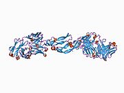
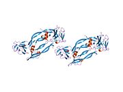
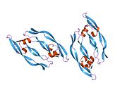
 1bj1: VASCULAR ENDOTHELIAL GROWTH FACTOR IN COMPLEX WITH A NEUTRALIZING ANTIBODY
 -
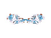
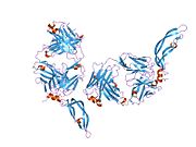
 1cz8: VASCULAR ENDOTHELIAL GROWTH FACTOR IN COMPLEX WITH AN AFFINITY MATURED ANTIBODY
 -
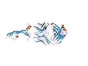
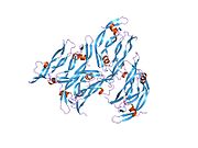
 1flt: VEGF IN COMPLEX WITH DOMAIN 2 OF THE FLT-1 RECEPTOR
 -
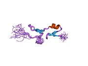
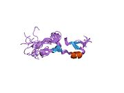
 1kat: Solution Structure of a Phage-Derived Peptide Antagonist in Complex with Vascular Endothelial Growth Factor
 -
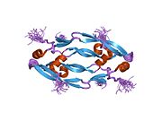
 1kmx: Heparin-binding Domain from Vascular Endothelial Growth Factor
 -
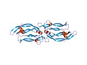
 1mjv: DISULFIDE DEFICIENT MUTANT OF VASCULAR ENDOTHELIAL GROWTH FACTOR A (C51A and C60A)
 -
 1mkg: DISULFIDE DEFICIENT MUTANT OF VASCULAR ENDOTHELIAL GROWTH FACTOR A (C57A and C102A)
 -
 1mkk: Disulfide deficient mutant of vascular endothelial growth factor A (C61A and C104A)
 -
 1qty: VASCULAR ENDOTHELIAL GROWTH FACTOR IN COMPLEX WITH DOMAIN 2 OF THE FLT-1 RECEPTOR
 -
 1tzh: Crystal Structure of the Fab YADS1 Complexed with h-VEGF
 -
 1tzi: Crystal Structure of the Fab YADS2 Complexed with h-VEGF
 -
 1vgh: HEPARIN-BINDING DOMAIN FROM VASCULAR ENDOTHELIAL GROWTH FACTOR, NMR, 20 STRUCTURES
 -
 1vpf: STRUCTURE OF HUMAN VASCULAR ENDOTHELIAL GROWTH FACTOR
 -
 1vpp: COMPLEX BETWEEN VEGF AND A RECEPTOR BLOCKING PEPTIDE
 -
 2fjg: Structure of the G6 Fab, a phage derived Fab fragment, in complex with VEGF
 -
 2fjh: Structure of the B20-4 Fab, a phage derived Fab fragment, in complex with VEGF
 -
 2vgh: HEPARIN-BINDING DOMAIN FROM VASCULAR ENDOTHELIAL GROWTH FACTOR, NMR, MINIMIZED AVERAGE STRUCTURE
 -
 2vpf: VASCULAR ENDOTHELIAL GROWTH FACTOR REFINED TO 1.93 ANGSTROMS RESOLUTION


